Microscopics: Or Why I Became a Medical Technologist
Photos and Commentary by Teri Stark
I cannot remember a time when I was not fascinated by what I could see with a microscope. As a child I wanted to be a doctor just so I could look in a scope all day long! I did not realize then that this was not their primary job. Fortunately for me I discovered Medical Technology. Here I am allowed to focus to my heart's content (or until the next STAT arrives!). For me, there is nothing like the feeling I get when I find that certain "thing" that will direct the doctor on the right path to curing his patient.
Granted, not every specimen is as exciting as the ones I am going to show you here, but every single specimen has that potential. Imagine a surprise in every urine container! Well, okay, maybe I do get a bit carried away with my enthusiasm. The microscopic world is an extraordinary adventure. Come with me while I show you a few things our lab has found in the past few years.
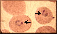 This photo is of malaria in a red blood cell (RBC). The arrows are pointing at the developing parasites. At this stage of development we call them malarial rings. As these parasites grow they cause the RBCs to burst. It is when these cells burst that the patient experiences chills. Ooh, cool, huh? Specimen source: whole blood. This photo is of malaria in a red blood cell (RBC). The arrows are pointing at the developing parasites. At this stage of development we call them malarial rings. As these parasites grow they cause the RBCs to burst. It is when these cells burst that the patient experiences chills. Ooh, cool, huh? Specimen source: whole blood.
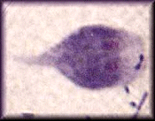 This little critter may look innocent but he is extremely unpleasant. His name is Giardia lamblia and he likes to live in warm, stagnant water. Colorado river water is one of his usual camping grounds. His nickname in the laboratory world is "Monkey Face". If you tilt your head to the right you can just barely make out his "face". This nasty little parasite will give you the worse case of intestinal disturbance you ever had. Not cool. Specimen source: feces. This little critter may look innocent but he is extremely unpleasant. His name is Giardia lamblia and he likes to live in warm, stagnant water. Colorado river water is one of his usual camping grounds. His nickname in the laboratory world is "Monkey Face". If you tilt your head to the right you can just barely make out his "face". This nasty little parasite will give you the worse case of intestinal disturbance you ever had. Not cool. Specimen source: feces.
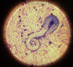 This worm is really, really, really not cool. Known as Strongyloides stercoralis, this parasite can reside for long periods of time in a human intestine and not cause a lick of problem. But get this fella excited and he will not make you happy! In this particular case the patient had been on medication that disturbed the worm. The worm (and a few of his close family members) migrated to the patient's lungs. They suspect the patient had been harboring the parasite since WWII when he was stationed in the Phillipines. This photo won
Photo of the Day for October 2, 1999 in the Macro category. Specimen source: lung tissue This worm is really, really, really not cool. Known as Strongyloides stercoralis, this parasite can reside for long periods of time in a human intestine and not cause a lick of problem. But get this fella excited and he will not make you happy! In this particular case the patient had been on medication that disturbed the worm. The worm (and a few of his close family members) migrated to the patient's lungs. They suspect the patient had been harboring the parasite since WWII when he was stationed in the Phillipines. This photo won
Photo of the Day for October 2, 1999 in the Macro category. Specimen source: lung tissue
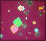 My most favorite thing to look at under the microscope! Who says urine is icky? These are uric acid crystals in urine. These crystals do not always indicate a pathogenic condition. They are sometimes present in normal patients as well as patients with gout. When I see them I immediately get out the polarizer attachment so I can see all the pretty colors. Specimen source: urine. My most favorite thing to look at under the microscope! Who says urine is icky? These are uric acid crystals in urine. These crystals do not always indicate a pathogenic condition. They are sometimes present in normal patients as well as patients with gout. When I see them I immediately get out the polarizer attachment so I can see all the pretty colors. Specimen source: urine.
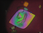 A closer look at a rainbow colored uric acid crystal from the same urine specimen. Most of the uric acid crystals will polarize to one color so this was a unique and pretty crystal. Not all urinary crystals will polarize light. We use the polarizer to distinguish between crystals that may be pathogenic but similar in shape. Specimen source: urine. A closer look at a rainbow colored uric acid crystal from the same urine specimen. Most of the uric acid crystals will polarize to one color so this was a unique and pretty crystal. Not all urinary crystals will polarize light. We use the polarizer to distinguish between crystals that may be pathogenic but similar in shape. Specimen source: urine.
The uric acid photomicrographs were taken with the Sony Cybershot S70 digital camera. The rest were taken with the Sony Digital Mavica model FD71. There were no special attachments for these shots. The cameras were held up to the eyepiece and the image captured. The wonders of the digital age! The photos were cropped, chopped, and framed using
Paint Shop Pro 7.
|


